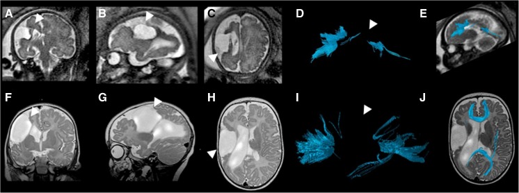
Fig. 5.
Fetal and postnatal corpus callosum segmentation in a boy (case 12) with right middle cerebral artery territory infarct. a–e Age 33+6 gestational weeks. a Coronal T2-weighted (TR/TE 2,042/140 ms, flip angle 90°) MR image of the brain reveals cystic encephalomalacia in the region of the right middle cerebral artery infarct (arrowhead). b Sagittal T2-weighted image of the fetal brain shows infarct (arrowhead). c Axial T2-weighted image of the fetal brain with infarct (arrowhead). d Sagittal view of the fiber tract segmentation reveals that the body of the corpus callosum is not visualized in the fetal diffusion tensor imaging (DTI; arrowhead). e Overlay of the callosal fiber tract on a sagittal T2-weighted image. f–j Age 4 months. f Coronal T2-weighted (TR/TE 3,000/120 ms, flip angle 90°) MR image of the brain shows sequelae of infarct (arrowhead). g Sagittal T2-weighted image shows infarct (arrowhead). h Axial T2-weighted image of the postnatal brain shows infarct (arrowhead). i Segmented sagittal DTI of the corpus callosum in the postnatal brain reveals that, similar to the fetal DTI, the body of the corpus callosum is not visualized. j An overlay of the segmented callosal fibers on an axial T2-weighted image reveals the markedly thinned parenchyma in the region of the right middle cerebral artery territory with cystic encephalomalacic changes. TE echo time, TR repetition time

