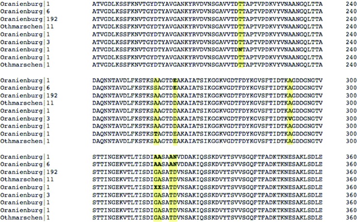
Fig. 4.
Multiple sequence alignment of Oranienburg and Othmarschen FliC protein sequences with conserved ends removed. Amino acid variants are in bold and highlighted in yellow. Sequence labels consist of the reported serovar and the number of times that unique FliC protein was seen within that serovar.

