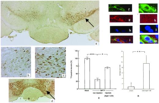
Figure 4.
(a) Photomicrographs of TH immunostaining in the substantia nigra of an MPTP-treated AAV-Apaf-1-DN-injected (arrow) mouse. (b) The noninjected side shows neuronal loss. (c) The injected side shows nos. of neurons compared with the noninjected side. (d) Photomicrographs of TH immunostaining in the substantia nigra of an AAV-EGFP-injected mouse (control infection) (arrow). Ratio of TH-positive cells between the noninjected and injected sides. The total number of TH-positive neurons was counted in three sections each from four different mice. Statistical analysis was performed by using ANOVA followed by Scheffé's post hoc test. Laser confocal image of TH immunostaining on the substantia nigra at the noninjected side (f) and the injected side (l). Laser confocal image of antiactivated caspase-3 staining on the substantia nigra at the noninjected side (g) and the injected side (k). DNA condensation can be detected by Hoechst 33258 staining. (h) Hoechst 33258 staining reveals an intact nucleus (l). Superimposed image (j and m). (n) Graph demonstrating the mean number of amphetamine-induced full body turns per minute. Left bar, MPTP-treated mice with injection of the vehicle; Right bar, MPTP-treated mice with injection of rAAV-Apaf-1-DN injection. **, P < 0.01. (a, ×20; b and c, ×100; d, ×10; f–m, ×600.)

