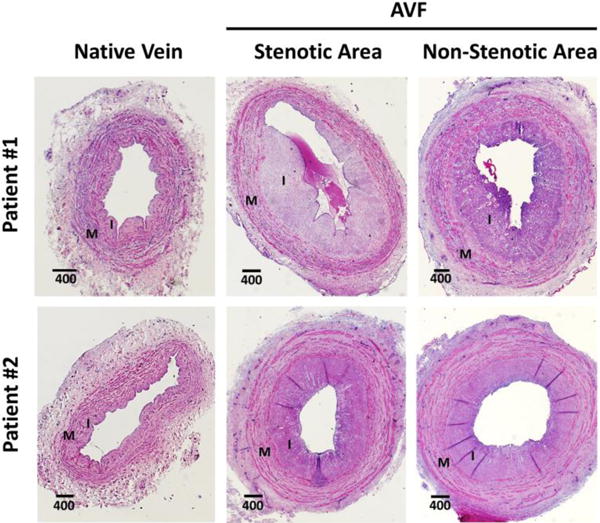
Figure 2.

Representative hematoxylin and eosin-stained cross-sections of native veins (left panel) before anastomosis and segments of the resulting arteriovenous fistulas (AVF; right panel) at the time of transposition. Native veins present a low-moderate degree of pre-existing intimal hyperplasia (IH), whereas AVFs show moderate-severe postoperative IH. Focal stenotic and nearby non-stenotic segments from the same AVF present a similar degree of IH. I: intima, M: media. Distances are in μm.
