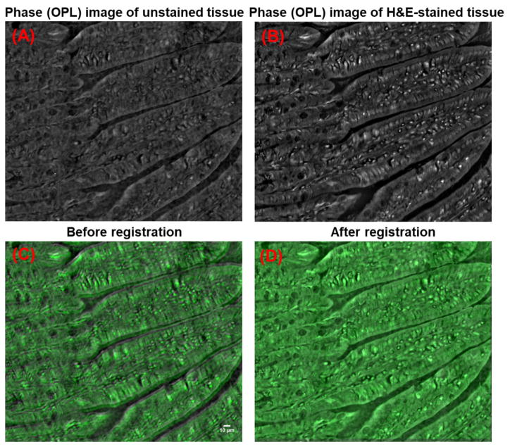
Figure 11.
Image registration based on the transmission phase (OPL) images of unstained and H&E-stained tissue. The transmission phase (OPL) images of (A) unstained and (B) stained tissue, as well as (C–D) the overlaid transmission phase images (gray: unstained tissue; green: H&E-stained tissue) (C) before and (D) after image registration.

