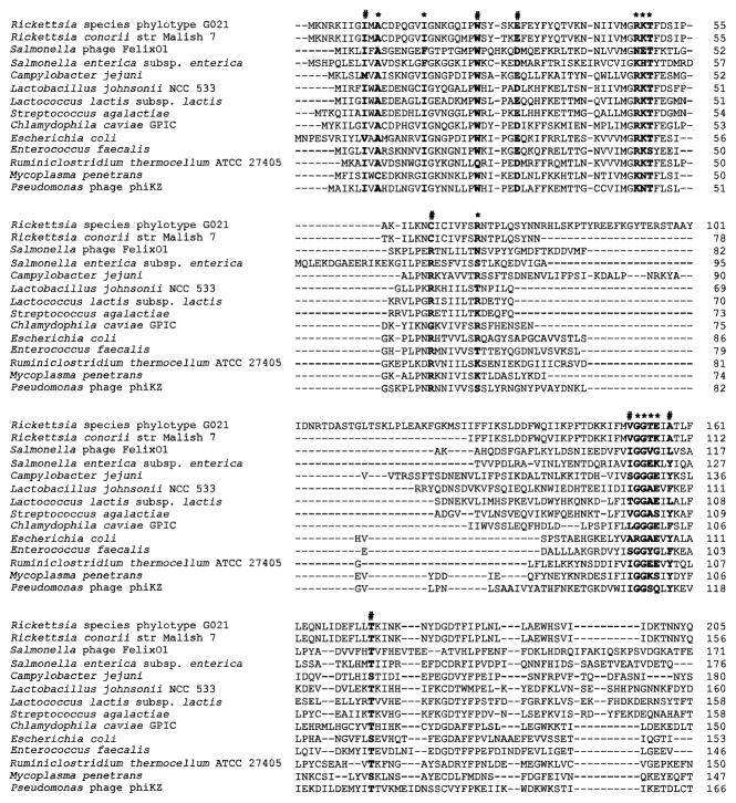
Figure 1.
Amino acid sequence alignment for dihydrofolate reductase (DHFR) from Rickettsia species phylotype G021 and other bacteria as well as phage species, including Campylobacter jejuni (GenBank accession number CAC19929), Chlamydophila caviae GPIC (AAP05737), Ruminiclostridium thermocellum ATCC 27405 (ABN52458), Enterococcus faecalis (WP_049097722), Escherichia coli (WP_063844334), Lactobacillus johnsonii NCC 533 (WP_011162224), Lactococcus lactis subsp. Lactis (EHE92570), Mycoplasma penetrans (WP_011077508), Pseudomonas phage phiKZ (NP_803570), Rickettsia conorii str Malish 7 (WP_010976722), Salmonella enterica subsp. Enterica (WP_032490189), Salmonella phage FelixO1 (NP_944929), and Streptococcus agalactiae (ODG94437). The first 205 (of 213) amino acids of Rickettsia species phylotype G021 FolA protein, and the corresponding amino acids from the other species listed, are shown. Residues for NADPH and substrate binding sites in DHFR enzymes are labeled with asterisks. The folate binding sites are noted with hashtags.

