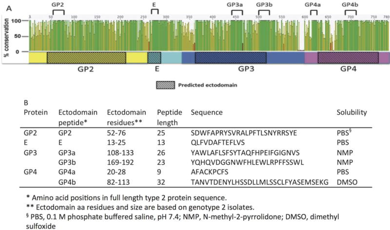
Figure 5.
Conserved PRRSV minor envelope ectodomain peptides. (A) Alignment of 2 type 1 and 56 type 2 PRRSV sequences. Open reading frames 2, 2b, 3, and 4 sequences were concatenated and translated to yield GP2 (yellow), E (aqua), GP3 (blue), and GP4 (pink) as shown. Sequence conservation is expressed as a percentage, where 100% indicates amino acid sites are completely conserved. Predicted ectodomain regions are marked with hashed boxes. Ectodomain peptide sequences with maximal conservation selected are marked by black lines at top of the figure. (B) Conserved ectodomain peptide position, length and sequences. The solvent used for initial dilution to 1mg/ml is indicated for each peptide.

