
Figure 1.
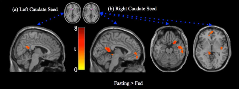
Paired t-test (fasting > fed) for caudate seed region connectivity. (a) Left caudate seed region connectivity is more strongly associated with activity in the posterior cingulate/precuneus in the fasting state (tmin = 2.9, tmax = 4.9). (b) Right caudate seed region connectivity is more strongly associated with the posterior cingulate cortex, medial prefrontal cortex, anterior insula and parahippocampus in the fasting state (tmin = 2.9, tmax = 6.2). There were no regions with stronger connectivity in the fed state.