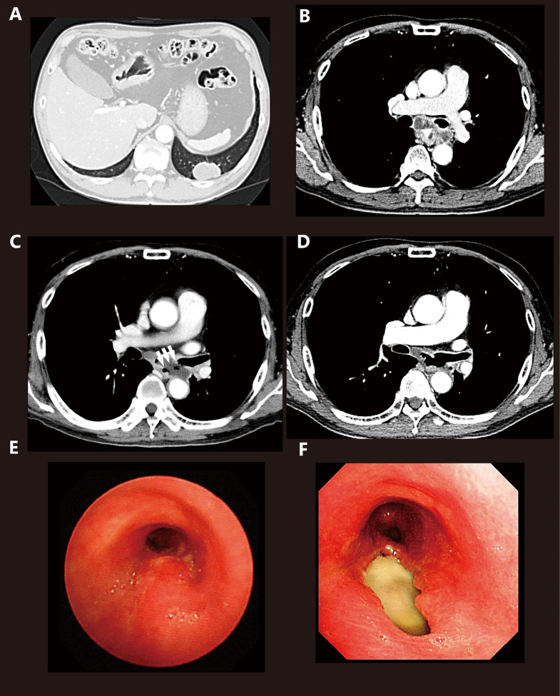
Fig. 1.
Computed tomography (CT) and bronchoscopy
(A) Chest CT showing the primary region in the left S9.
(B) Chest CT showing the subcarinal lymph node compressing the esophagus before treatment and the subcarinal lymph node exerting pressure on the left main bronchus (arrow head).
(C) A tracheoesophageal fistula was observed between the left main bronchus and esophagus through the subcarinal lymph node (arrows).
(D) Chest CT performed 2 weeks after diagnosis of TEF showing a remarkably diminished subcarinal lymph node enlargement after chemotherapy.
(E) An elevated membranous portion of the left main bronchus observed by bronchoscopy before treatment.
(F) The fistula (2.5 cm × 1.0 cm) was observed in the left main bronchus.

