Figure 4.
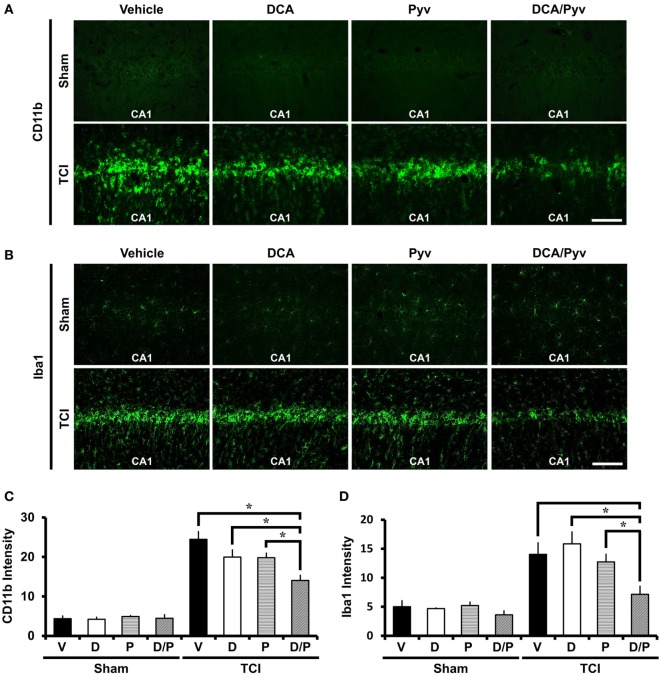
Combined treatment with dichloroacetic acid (DCA) and pyruvate reduces transient cerebral ischemia (TCI)-induced microglia activation. TCI-induced microglial activation was reduced by the co-treatment of DCA and pyruvate. (A) Conjugated donkey anti-mouse (CD11b) fluorescence microscopic images in the cornus ammonis 1 (CA1) region, which shows microglia and macrophage activation in the TCI-induced groups. CD11b expressed fluorescence signal was reduced in the combined treatment of the DCA and pyruvate group, compared to the vehicle-treated group. Scale bar = 100 μm (Normal-vehicle, n = 5; Normal-DCA, n = 5; Normal-pyruvate, n = 5; Normal-DCA + pyruvate, n = 5; TCI-vehicle, n = 5; TCI-DCA, n = 5; TCI-pyruvate, n = 7; TCI-DCA + pyruvate, n = 7). (B) Iba1 fluorescence microscopic images in the hippocampal CA1, which shows specific expression in microglia within the brain (Normal-vehicle, n = 5; Normal-DCA, n = 5; Normal-pyruvate, n = 5; Normal-DCA + pyruvate, n = 5; TCI-vehicle, n = 5; TCI-DCA, n = 5; TCI-pyruvate, n = 6; TCI-DCA + pyruvate, n = 5). (C,D) The bar graph represents the intensity of activated CD11b and Iba1 in the CA1 area that was based on CD11b and Iba1 fluorescence signal. Data are mean ± SEM, n = 5–7 from each group. D, DCA; P, pyruvate; D/P, DCA + pyruvate; V, vehicle.

