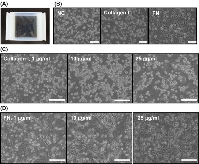
Figure 1. Silicone elastomer chamber and optimization of extracellular matrix (ECM) coating by morphological observation.
(A) A top view of the silicone-based elastomer chamber. (B) Morphology of rat aortic smooth muscle cells (SMCs) seeded on noncoated (NC), type I collagen, and fibronectin (FN)-coated and silicone chambers. ECM precoating was performed by incubation with either PBS, 25 μg/ml of type I collagen, or 10 μg/ml of FN at 24 h before seeding. Microphotographs were taken at 60 min after seeding. Optimization of ECM coating dose was determined by seeding 8 × 10⁵ of SMCs onto the silicone chambers precoated with indicated doses of type I collagen (C) or FN (D). Cell morphology observed at 60 min after seeding showed that FN exerted better cell spreading effect on SMCs; scale bars = 20 μm.

