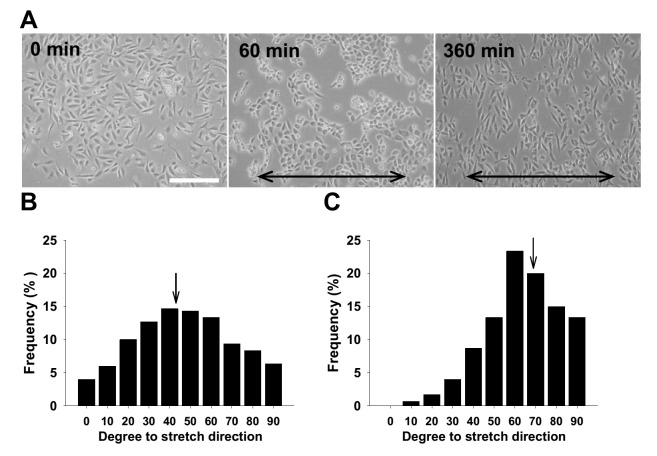
Figure 3. Reorientation and alignment of rat aortic smooth muscle cells (SMCs) induced by cyclic mechanical stretch.
(A) About 8 × 10⁵ of SMCs were seeded onto fibronectin-coated (at 10 μg/ml) silicone elastomer chambers for 3 h till cellular attachment and spreading, followed by consecutively uniaxial and cyclic 10% deformation at constant frequency (1 Hz). The morphology was observed and documented under inverted microscope at 0, 60, and 360 min. The lines with arrows on both ends indicate stretching direction; scale bars = 20 μm. Morphometrics was used to measure the angles between SMC long axes and stretching direction. Representative angle histograms of SMCs before (B) and after 360 min stretching (C) are shown. Arrows indicate the mean angle locations, 42.5 and 67.9 degrees respectively.

