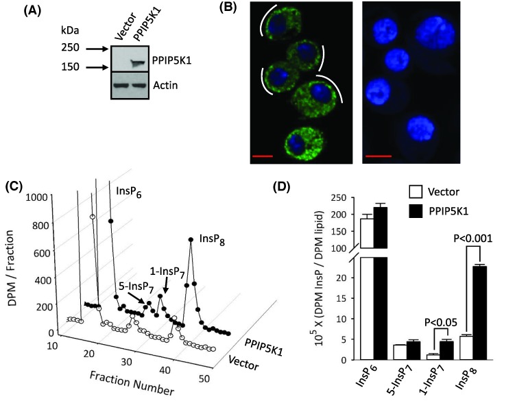
Figure 2. The impact of heterologous expression of PPIP5K1 upon PP-InsP turnover in Drosophila S3 cells.
(A) Western analysis of PPIP5K1 expression in S3vector and S3PPIP5K1 cells. (B) Confocal immunofluorescence analysis of PPIP5K1 distribution and Hoechst staining in S3PPIP5K1 cells (left panel) or S3vector cells (right panel); white curved lines highlight a tendency for the PPIP5K1 to be more concentrated in punctae near the plasma membrane; scale bars = 5 μm. (C) HPLC chromatograph of InsP6 and PP-InsPs in [3H]inositol-labeled S3vector and S3PPIP5K1 cells. (D) Cellular levels of InsP6 and PP-InsPs, normalized to levels of [3H]inositol-lipids, from three independent experiments similar to that described in panel C.

