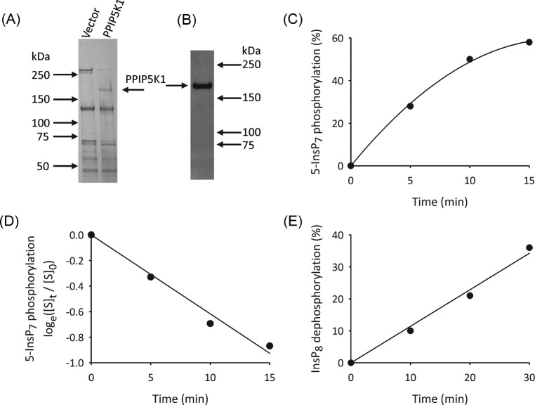
Figure 3. Analysis of recombinant PPIP5K1.
(A) Silver-stained, SDS/PAGE analysis of recombinant PPIP5K1 purified from S3PPIP5K1 cells, compared with S3vector cells. (B) Western analysis of recombinant PPIP5K1. (C) Representative time course of 5-InsP7 phosphorylation by 30 ng PPIP5K1 and just trace amounts of [3H]-labeled substrate (i.e under first-order conditions [24]). (D) Natural logarithmic transformation of the data in panel C, where [S]0 = [3H]substrate at time zero, and [S]t = [3H]substrate at time t, as indicated. (E) Time course of InsP8 dephosphorylation by 220 ng recombinant PPIP5K1, determined by HPLC.

