Figure 1.
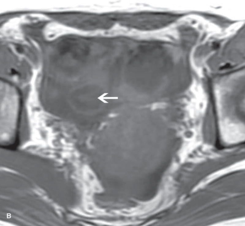
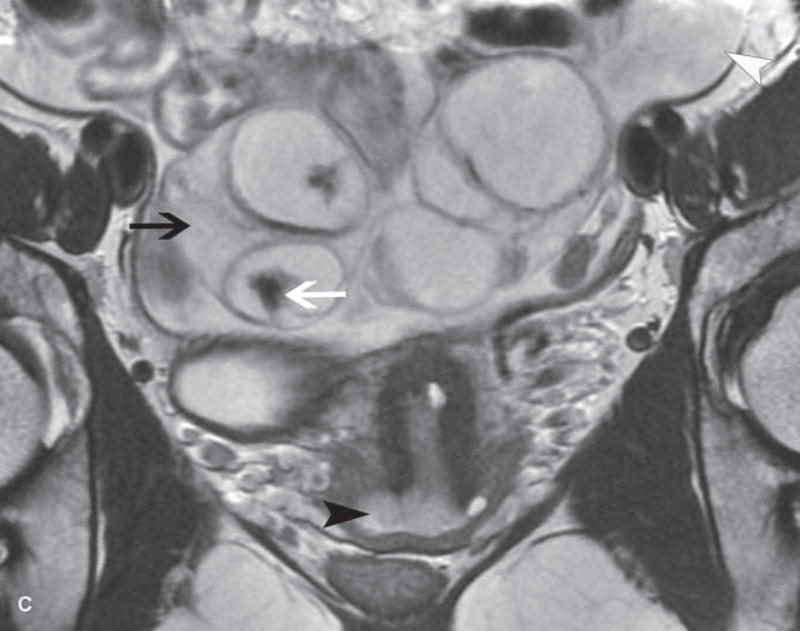
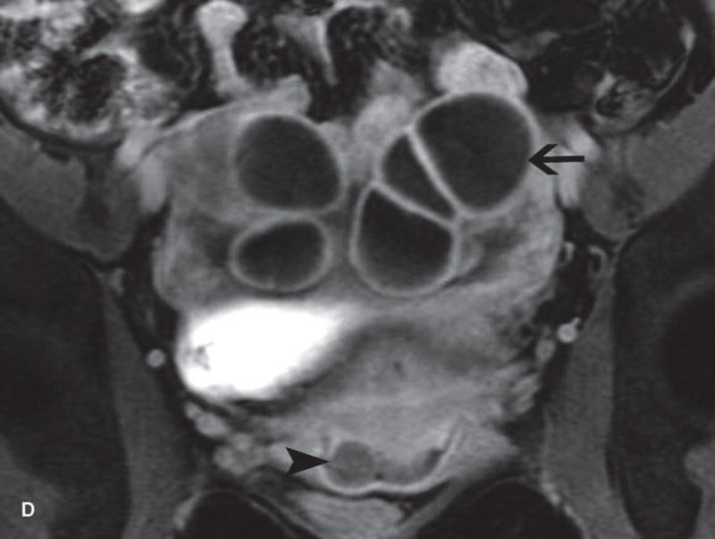
31-year-old woman with cervical cancer and ovarian hyperstimulation. A. A drawing shows the spoke-wheel appearance of hyperstimulated ovaries with multiple peripheral cysts centered on ovarian stroma. B. Axial T1WI shows symmetrically enlarged bilateral ovaries with low SI cysts; some cysts contain high SI blood products (white arrow). C. Axial oblique T2WI shows symmetrically enlarged bilateral ovaries with high SI cysts peripherally and edematous stroma centrally (black arrow); some cysts contain low SI foci due to blood products (white arrow). Note the small volume ascites (white arrowhead) and primary cervical tumor (black arrowhead). D. Axial oblique contrast-enhanced image shows symmetrically enlarged ovaries. Ovaries contain peripheral cysts (black arrow) with wall enhancement and enhancing central stroma. Note the absence of enhancing nodular septa or nodules. Primary cervical tumor is also seen (black arrowhead).


