Figure 5.
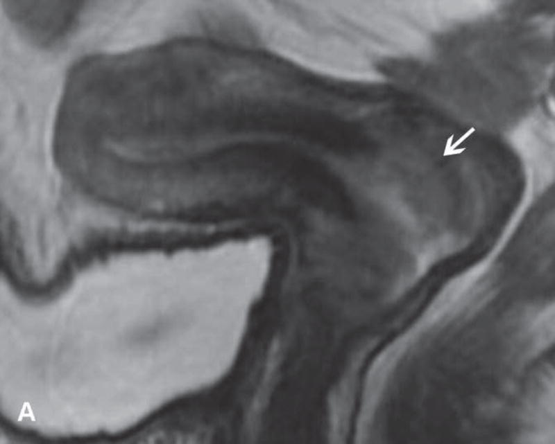
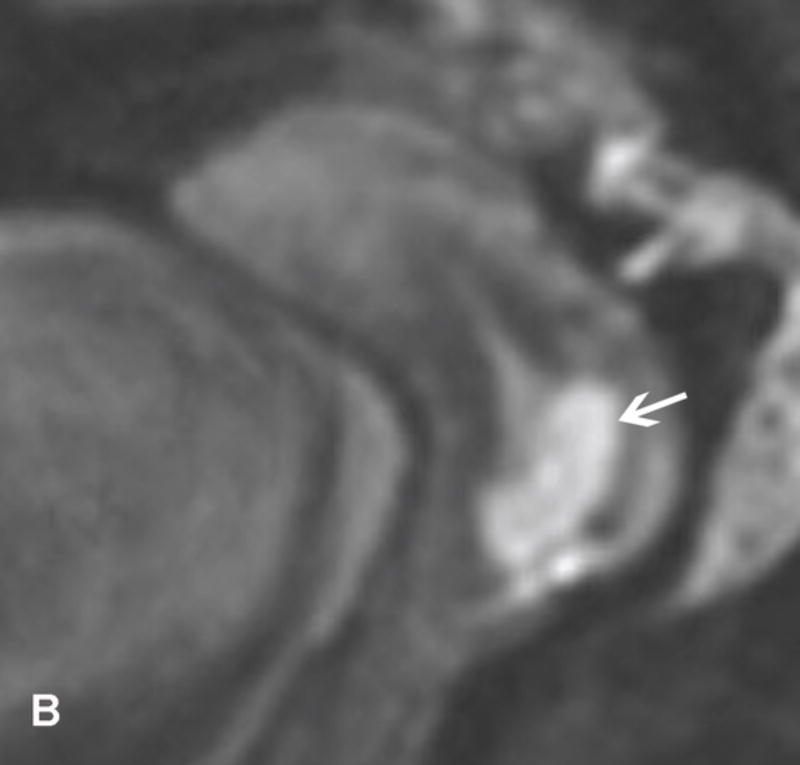
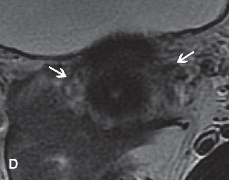
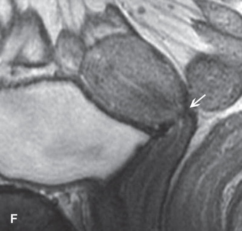
31-year-old woman with Stage IB1 cervical cancer. A. Sagittal T2WI demonstrates endophytic intermediate-SI tumor (white arrow). The tumor borders are obscured by post-cone resection changes making it challenging to accurately determine tumor size and endocervical extent. B and C. The tumor margins (white arrow) are more clearly defined on DWI and contrast-enhanced images, respectively, facilitating assessment of tumor size (2 cm in maximum dimension) and extent (1 cm caudal to internal os). D and E. Axial oblique and coronal oblique T2WI illustrate the location of internal os marked by the entrance of the uterine vessels (white arrows). F. Sagittal T2WI in the same patient after radical trachelectomy shows the normal appearing uterine remnant and isthmic vaginal anastomosis (white arrow).


