
Figure 6.
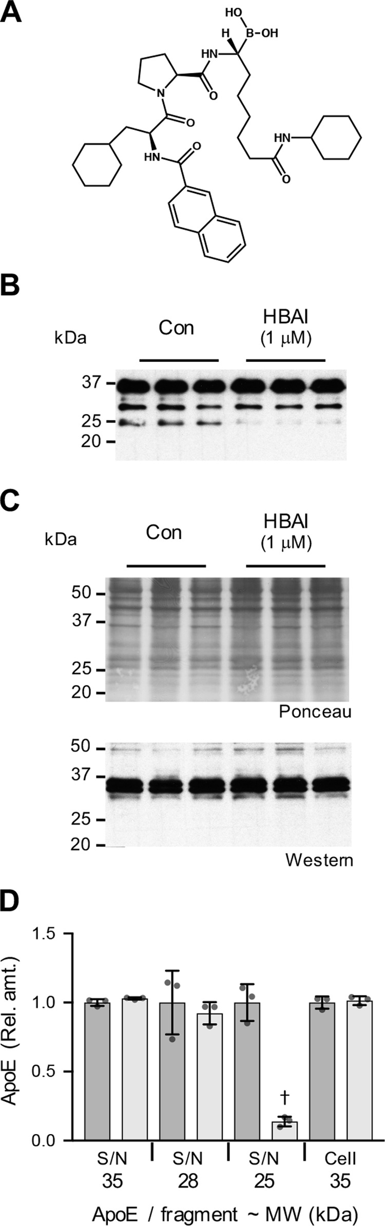
HtrA1 inhibitor reduces SK-N-SH cellular apoE proteolytic fragmentation. The structure of HBAI is shown (A). SK-N-SH cells were cultured for 9 days in the presence of 10 μm ATRA. The culture medium was then replaced with fresh medium alone (Con) or with fresh medium containing either HBAI (1 μm) or vehicle control and cultured for a further 24-h period. The addition of HBAI inhibited the formation of the apoE 25-kDa fragment in the supernatant (S/N) (B). HBAI addition had no impact on cellular apoE levels (C). Quantitative assessments of the full-length apoE (35 kDa) and apoE fragments at 28 and 25 kDa indicate a significant inhibition of apoE 25-kDa fragment formation in the presence of HBAI (light gray bars; D). Data in D are relative optical density measurements in which the vehicle control samples (DMSO (1:1,000), dark gray bars) are defined as 1.0. Data in D are derived from one experiment performed in triplicate (Western blots shown in B and C) and are representative of three independently performed experiments. The histogram bars represent mean values, and the error bars represent S.D. †, p < 0.01 compared with the respective control for each fragment; two-tailed t test.