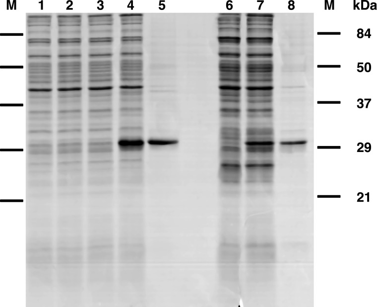
Figure 1.
Expression in Escherichia coli and purification of AtUCP1 and AtUCP2. Proteins were separated by SDS-PAGE and stained with Coomassie Blue. Lanes 1–5, AtUCP1; lanes 6–8, AtUCP2. Markers were Bio-Rad prestained SDS-PAGE standards: bovine serum albumin, 84 kDa; ovalbumin, 50 kDa; carbonic anhydrase, 37 kDa; soybean trypsin inhibitor, 29 kDa; lysozyme, 21 kDa). Lanes 1–4, E. coli BL21(DE3); lanes 6 and 7, E. coli BL21 CodonPlus(DE3)-RIL containing the expression vector, without (lanes 1, 3, and 6) and with the coding sequence of AtUCP1 (lanes 2 and 4) and the coding sequence of AtUCP2 (lane 7). Samples were taken immediately before induction (lanes 1 and 2) and 5 h later (lanes 3, 4, 6, and 7). The same number of bacteria were analyzed in each sample. Lanes 5 and 8, purified AtUCP1 protein (5 μg) and purified AtUCP2 (3 μg) derived from bacteria shown in lanes 4 and 7, respectively.

