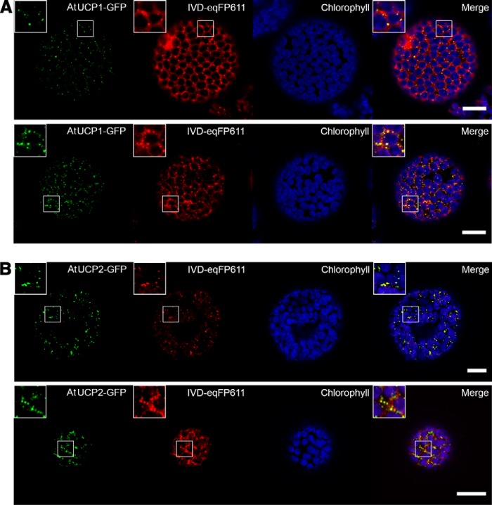
Figure 8.
Subcellular localization of AtUCP1-/AtUCP2-GFP fusion proteins in N. benthamiana protoplasts. Fluorescent signals of AtUCP1-/AtUCP2-GFP (green), mitochondrial marker IVD-eqFP611 (red), chlorophyll A/chloroplasts (blue), and merge showing the overlap of the fluorescent signals (yellow) detected via confocal laser-scanning microscopy. A, co-localization of AtUCP1-GFP with the mitochondrial marker. B, co-localization of AtUCP2-GFP with the mitochondrial marker. Scale bar, 20 μm. Two independently transformed cells are shown in each panel.

