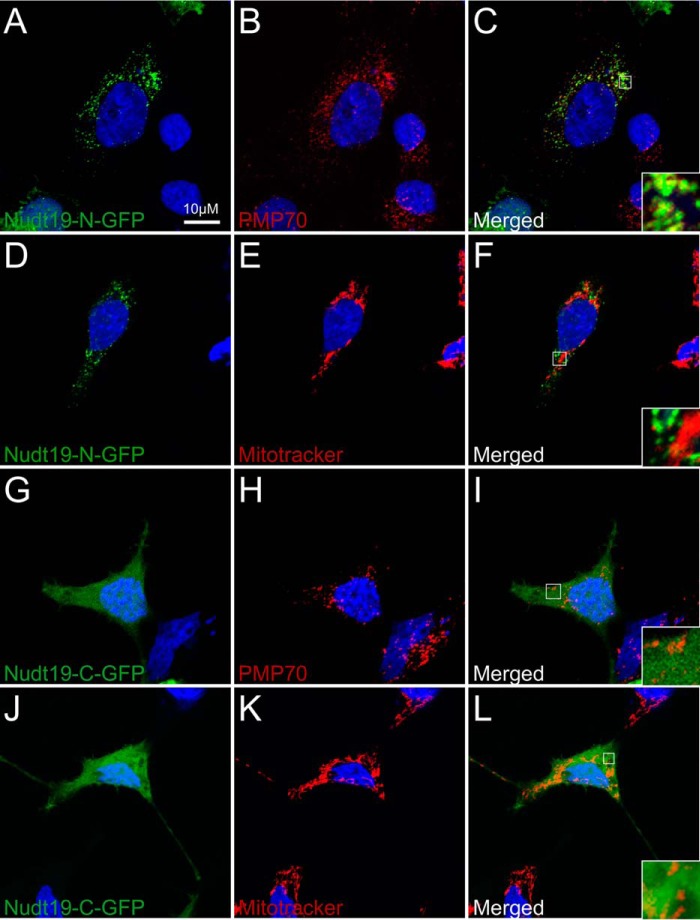
Figure 3.
Localization of Nudt19 to the peroxisomes. HEK 293 cells were transfected with expression plasmids encoding for GFP fused to the N terminus (Nudt19-N-GFP) (A–F) or the C terminus of Nudt19 (Nudt19-C-GFP) (G–L). Fixed cells were visualized using confocal microscopy. B, C, H, and I, the endogenous PMP70 protein was used as a marker for the peroxisomes, shown in red. E, F, K, and L, mitochondria were visualized using MitoTracker Orange CMTMRos and shown in red. A–L, cell nuclei were stained with DAPI, in blue. C, F, I, and L, insets in merged images show details at higher magnification. The results are representative of at least two independent experiments.

