
Figure 3.
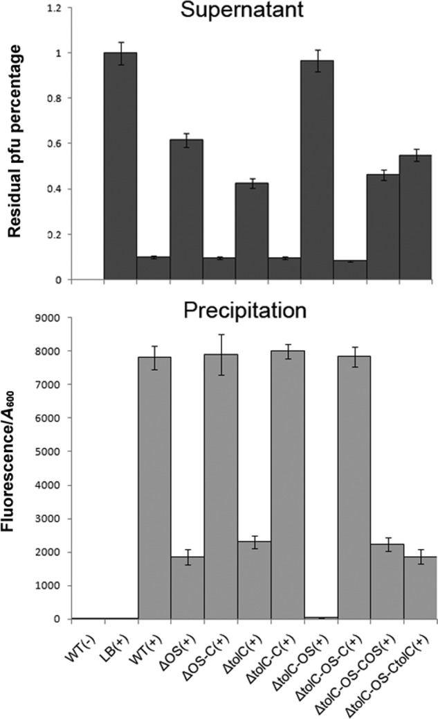
Analysis of the binding capacity between isolates and VP3 phage. The VP3 phage (109 cfu/ml) was labeled with SYBR Gold and mixed with fresh N16961 culture (A600 = 0.1) in a 1:1 (v/v) ratio for 5 min, and each sample was centrifuged at 4,000 rpm for 5 min. The remaining bacterial precipitation was resuspended in 200 μl of SM, and the fluorescence value was determined at 490 nm excitation/537 nm emission. Binding capacity between the strain and VP3 is measured via total fluorescence value/A600. The remaining phage titer in the supernatant of each sample was determined using a double-layer plaque assay. LB culture medium containing only wildtype strain or VP3 phage was used as a negative control, and the phage titer in the control supernatant was set to 100%. Error bars indicate ranges. +, VP3 present; −, VP3 not present, the detailed information of strains in Table 2.