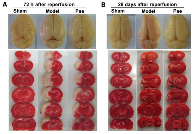
Figure 2.
Effects of paeonol on ischemic brain injury. Representative images of total brains indicated brain lesions at (A) 72 h and (B) 28 days after reperfusion (upper panel). Representative images of TTC-stained coronal slices with 2 mm-thickness indicated ischemic lesions at (A) 72 h and (B) 28 days after reperfusion (lower panel). After TTC staining, the viable tissue stained deep red, whereas the infarct area was white in color. TTC, 2,3,5-triphenyltetrazolium chloride; Pae, paeonol.

