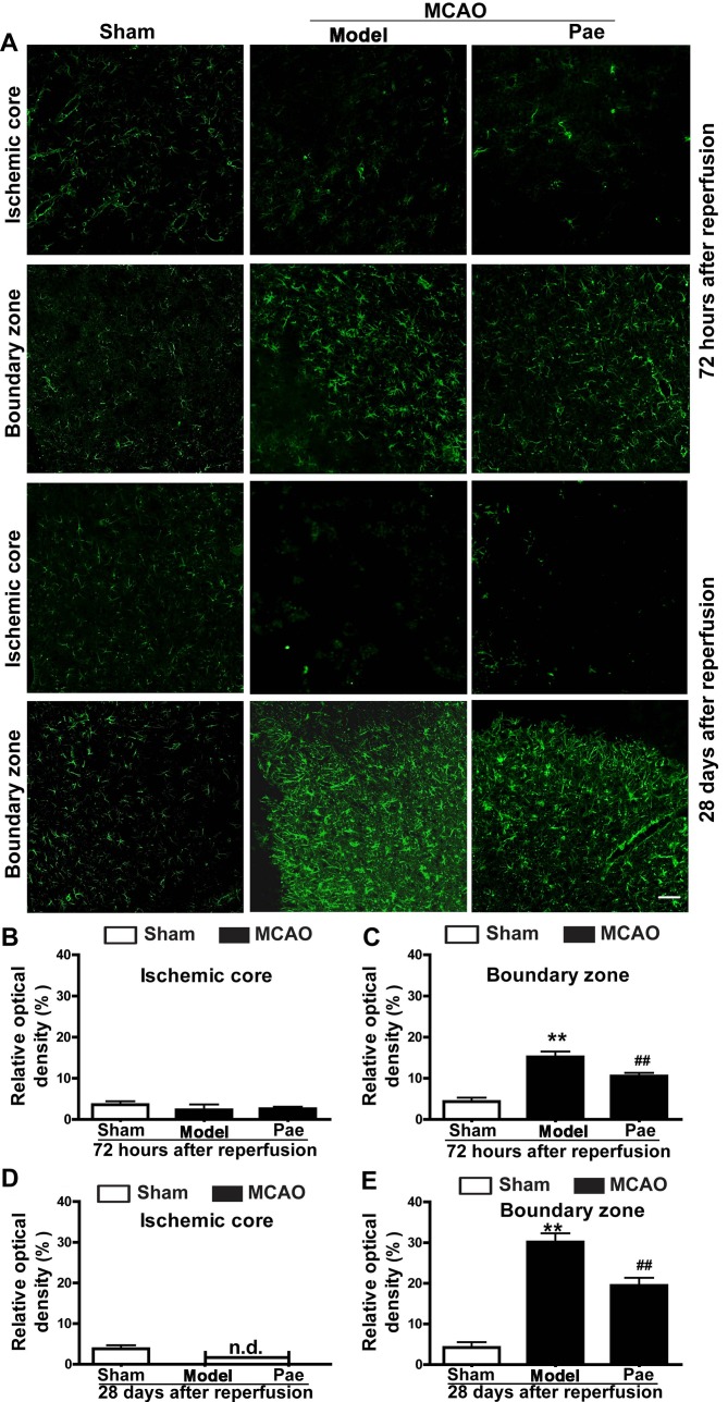
Figure 4.
Effects of paeonol on astrocyte proliferation at 72 h and 28 days after reperfusion in rats. (A) Representative photomicrographs indicating glial fibrillary acidic protein-immunopositive astrocytes at 72 h and 28 days after reperfusion (scale bar, 100 µm) and (B-E) quantification results. At 72 h after reperfusion, astrocytes were initially increased in the boundary zone, and paeonol treatment significantly suppressed the changes in the boundary zone, but not those in the ischemic core. At 28 days after reperfusion, astrocytes were significantly increased in the boundary zone, and paeonol treatment significantly attenuated the astrogliosis in the boundary zone, but not that in the ischemic core. Values are expressed as the mean ± standard error of the mean from eight rats per group. **P<0.01 compared with sham group, ##P<0.01 compared with model group. Pae, paeonol; MCAO, middle cerebral artery occlusion; n.d., not detectable.

