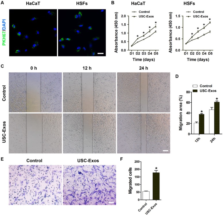
Figure 2.
USC-Exos enhance the proliferation and migration of keratinocytes and fibroblasts. (A) Fluorescence microscopy analysis of PKH67-labeled USC-Exos internalization by human keratinocytes cell line HaCaT and skin fibroblasts (HSFs). The green-labeled exosomes were visible in the perinuclear region of recipient cells. Scale bar: 50 μm. (B) The proliferation of HaCaT and HSFs receiving different treatments was tested by CCK-8 analysis. n = 4 per group. (C) Representative images of scratch wound assay in HaCaT treated with USC-Exos or PBS. Scale bar: 250 μm. (D) Quantitative analysis of the migration rates in (C). n = 3 per group. (E) The migration of HSFs stimulated by USC-Exos or an equal volume of PBS was detected by the transwell assay. Scale bar: 100 μm. (F) Quantitative analysis of the migrated cells in (E). n = 3 per group. *P < 0.05 vs. PBS (control) group.

