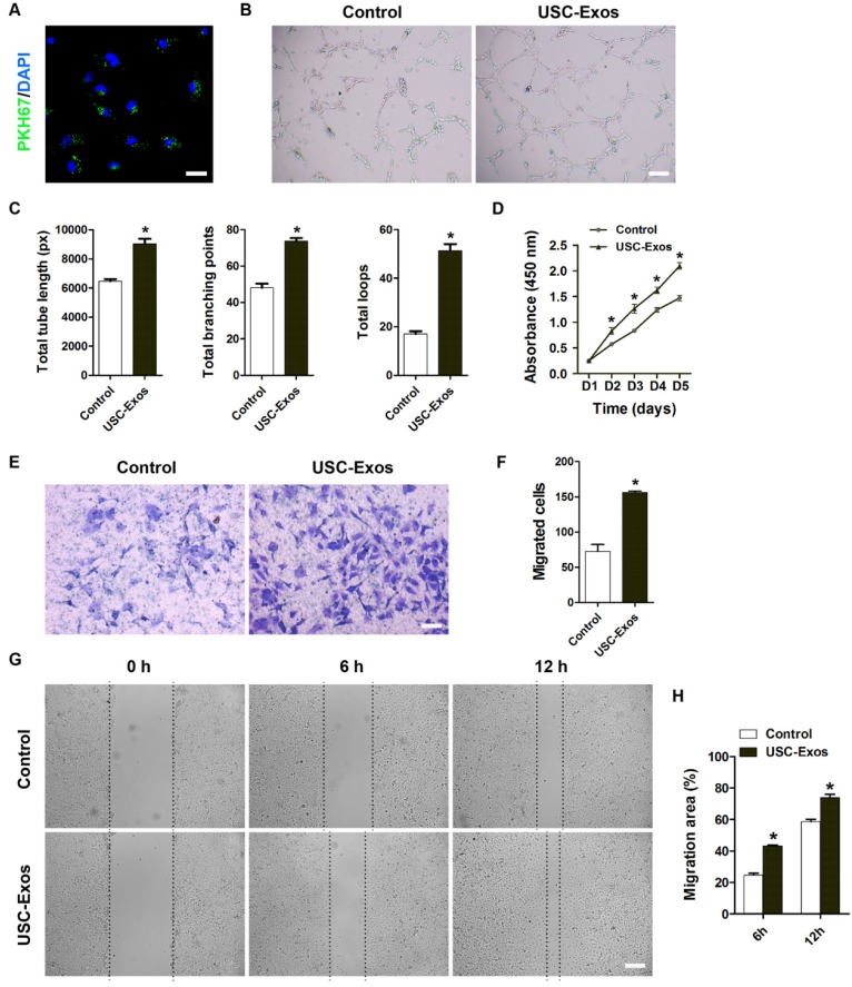
Figure 3.
USC-Exos augment the angiogenic activities of endothelial cells. (A) Fluorescence microscopy analysis revealed that PKH67-labeled USC-Exos were incorporated into human microvascular endothelial cells (HMECs). Scale bar: 50 μm. (B) Representative images of the tube formation assay on Matrigel in HMECs treated with USC-Exos or PBS. Scale bar: 200 μm. (C) Quantitative analyses of the total tube length, total branching points and total loops in (B). n = 3 per group. (D) HMECs exhibited a much stronger proliferative ability when exposed to USC-Exos, as tested by CCK-8 analysis. n = 4 per group. Transwell assay (E-F) and scratch wound healing assay (G-H) revealed that USC-Exos up-regulated the motility of HMECs. Scale bar: 100 μm (E) or 250 μm (G). n = 3 per group. *P < 0.05 vs. PBS (control) group.

