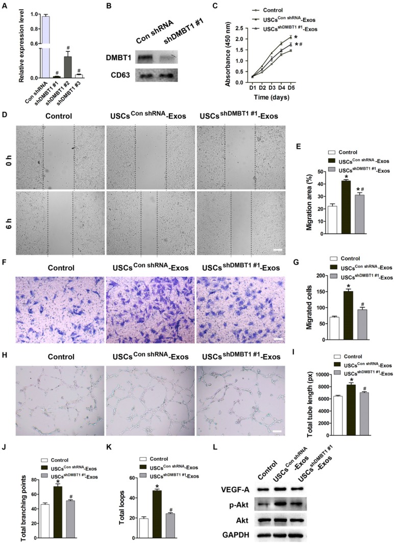
Figure 5.
DMBT1 mediates the pro-angiogenic effects of USC-Exos on endothelial cells. (A) The inhibitory efficiency of shRNAs targeting DMBT1 was verified by qRT-PCR analysis. n = 3 per group. #P < 0.05 vs. Con shRNA group. (B) Western blot analysis of DMBT1 in exosomes from DMBT1-silenced USCs (USCsshDMBT1 #1-Exos) and from Con shRNA-treated USCs (USCsCon shRNA-Exos). (C) The proliferation of HMECs treated with PBS, USCsCon shRNA-Exos and USCsshDMBT1 #1-Exos was tested by CCK-8 analysis. n = 4 per group. (D-G) The migration of HMECs stimulated by PBS, USCsCon shRNA-Exos and USCsshDMBT1 #1-Exos was detected by the scratch wound assay (D-E) (Scale bar: 250 μm) and the transwell assay (F-G) (Scale bar: 100 μm). n = 3 per group. (H) Representative images of the tube formation assay in HMECs treated with PBS, USCsCon shRNA-Exos and USCsshDMBT1 #1-Exos. Scale bar: 200 μm. (I-K) Quantitative analyses of the total tube length, total branching points and total loops in (H). n = 3 per group. (L) Detection of the protein levels of VEGF-A, Akt and p-Akt in HMECs receiving different treatments by western blotting. n = 3 per group. *P < 0.05 vs. PBS (control) group, #P < 0.05 vs. USCsCon shRNA-Exos group.

