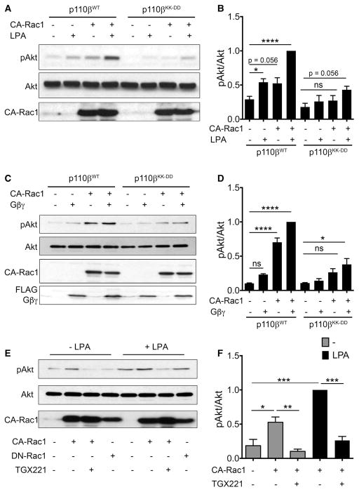
Figure 1. CA-Rac1 does not activate GPCR-uncoupled p110β.
(A) MDA-MB-231 cells stably expressing wild-type or GPCR uncoupled p110β (p110βKK-DD) were transiently transfected with control plasmid or GFP-CA-Rac1, and stimulated with or without 10 μM LPA for 5 min. Representative immunoblots show pT308-Akt, total Akt, and GFP-CA-Rac1. (B) Quantitation of pAkt/Akt ratios from immunoblots in (A). pAkt/Akt ratios in each experiment were normalized to that seen in LPA-stimulated cells expressing wild-type p110β and GFP-CA-Rac1. The data represent the mean ± SEM from four independent experiments. (C) MDA-MB-231 cells stably expressing wild-type p110β or p110βKK-DD were transiently transfected with control plasmid, GFP-CA-Rac1, and/or Flag-Gβγ plasmids. Representative immunoblots show pT308-Akt, total Akt, GFP-CA-Rac1, and Flag-Gβγ. (D) Quantitation of pAkt/Akt ratios from immunoblots in (C). pAkt/Akt ratios in each experiment were normalized to that seen in cells simultaneously expressing wild-type p110β, GFP-CA-Rac1, and Gβγ. The data represent the mean ± SD from two independent experiments. (E) Parental MDA-MB-231 cells were transiently transfected with GFP-CA-Rac1 or GFP-DN-Rac1, and stimulated with LPA and treated with TGX-221 as indicated. Representative immunoblots show pT308-Akt, total Akt, and GFP-CA-Rac1. (F) Quantitation of pAkt/Akt ratios from (E) pAkt/Akt ratios in each experiment were normalized to that seen in cells expressing GFP-CA-Rac1 without TGX-221 treatment. Quantitation of the DN-Rac1 data in (E) is shown in Figure 3A. *P < 0.05; **P < 0.01; ****P < 0.0001; ns: not significant.

