Figure 2.
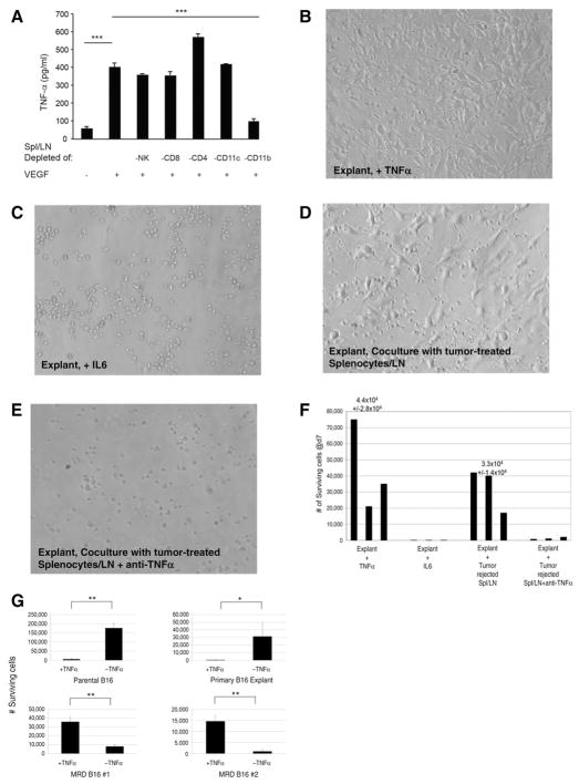
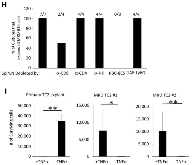
MRD cells use TNFα as a growth factor. A, A total of 105 splenocytes and lymph node cells from C57BL/6 mice cleared of B16tk tumors (after ganciclovir) were depleted of asialo GM-1+(NKs), CD4+, CD8+, CD11c+, or CD11b+cells by magnetic bead depletion and plated in the presence or absence of VEGF165 (12 ng/mL) in triplicate. Cell supernatants were assayed for TNFα by ELISA after 48 hours. Mean and SD of triplicate wells are shown. Representative of two separate experiments.***, P < 0.0001 (t test). B and C, Skin from the B16tk cell injection site from mice treated with ganciclovir (no palpable tumor after regression) was treated with TNFα (B; 100 ng/mL) or IL6 (C; 100 pg/mL). Seven days later, wells were inspected for actively growing tumor cell cultures. Images are representative of 15 skin explants over five different experiments. D and E, Skin explants from the site of B16tk cell injection of mice treated with ganciclovir (no palpable tumor) cocultured with 105 splenocytes and lymph node cells from C57BL/6 mice cleared of B16tk tumors after ganciclovir treatment alone (D) or in the presence of anti-TNFα (E; 0.4 μg/mL). Seven days later, wells were inspected for actively growing tumor cells. Images are representative of 5 separate explants. F, Quantitation of B–E. Three separate explants per treatment were counted. G, A total of 104 parental B16 cells, explanted B16 cells from a PBS-treated mouse, or cells from two MRD B16 cultures (expanded in vitro in TNFα for 72 hours) were plated in triplicate and grown in the presence or absence of TNFα for 4 days. Surviving cells were counted. Mean and SD of triplicates are shown. Representative of three experiments. *, P < 0.01; **, P < 0.001 (ANOVA). H, Splenocytes and lymph node cells from C57BL/6 mice cleared of B16tk tumors after ganciclovir treatment were treated with no antibody or with depleting antibodies specific for CD8, CD4, asialo GM-1 (NK cells), monocytes and macrophages, or neutrophils. Skin samples from regressed tumor sites were cocultured with 105 depleted or nondepleted splenocytes and lymph node cultures in the presence of VEGF165 (12 ng/mL). Seven days later, wells were inspected for actively growing tumor cell cultures. The percentage of cultures positive for active MRD growth (wells contained >104 adherent B16 cells) is shown. I, A total of 104 explanted TC2 tumor cells from a PBS-treated mouse or cells from two MRD TC2 cultures (expanded in vitro in TNFα for 72 hours) were plated in triplicate and grown in the presence or absence of TNFα for 4 days. Surviving cells were counted. Mean and SD of triplicates are shown. *, P < 0.01; **, P < 0.001 (ANOVA).


