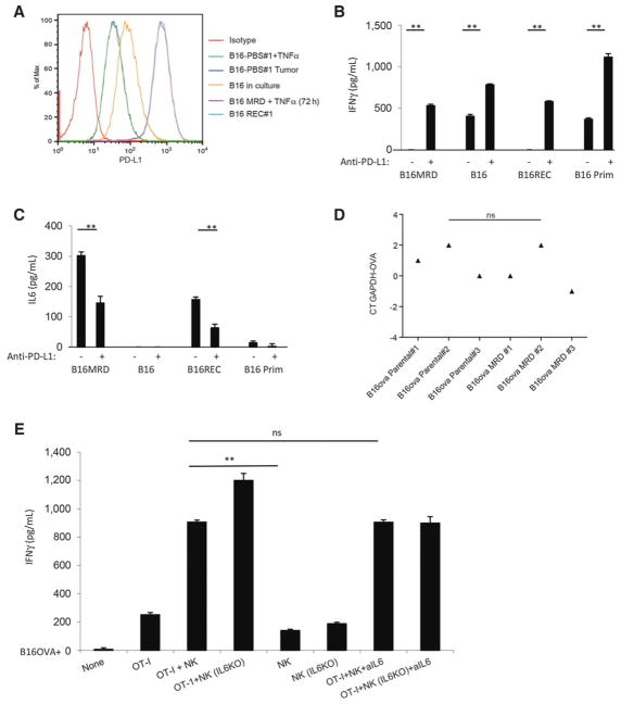
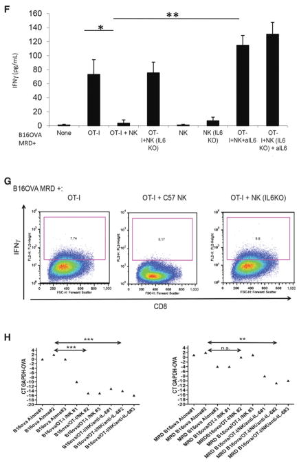
Figure 5.
PD-L1 expression on MRD inhibits immunosurveillance through IL6. A, Expression of PD-L1 was analyzed by flow cytometry on parental B16 cells in culture. Cells from a small (~0.3 cm diameter) B16tk tumor explanted from a PBS-treated mouse were cultured for 72 hours in vitro alone (B16-PBS#1; dark blue) or with TNFα (B16-PBS#1 + TNFα; green). B16 MRD cells recovered from the site of B16tk cell injection after regression were treated with TNFα for 72 hours (B16 MRD + TNFα 72 hours; purple). Cells from a small recurrent B16tk tumor (~0.3 cm diameter) explanted following regression after ganciclovir treatment was cultured for 72 hours without TNFα (B16 REC#1; light blue). Representative of three separate experiments. B and C, MRD B16 cells expanded for 72 hours in TNFα, parental B16 cells, explanted B16tk recurrent tumor cells, or explanted primary B16 tumors were plated (104 cells/well). Twenty-four hours later, 105 purified NK cells from C57BL/6 mice were added to the wells with control IgG or anti–PD-L1. Forty-eight hours later, supernatants were assayed for IFNγ (B) or IL6 (C) by ELISA. Means of triplicates per treatment are shown. Representative of three separate experiments (ANOVA). D, cDNA from three explants of PBS-treated B16ova primary tumors (~0.3 cm diameter) and three MRD B16ova cultures (derived from skin explants after regression with OT-I T-cell therapy and growth for 72 hours in TNFα) were screened by qRT-PCR for expression the ova gene. Relative quantities of ova mRNA were determined (ANOVA). Statistical significance was set at P < 0.05 for all experiments. E and F, A total of 104 parental B16ova cells (E) or MRD B16ova cells (F; derived as previously stated) were cocultured with purified CD8+ OT-I T cells and/or purified NK cells from either wild-type C57BL/6 or from IL6 KO mice (OT-I:NK:tumor 10:1:1) in triplicate in the presence or absence of anti-IL6. Seventy-two hours later, supernatants were assayed for IFNγ by ELISA. Mean and SD of the triplicates are shown. Representative of three separate experiments. **, P < 0.01 (ANOVA). G, A total of 104 B16ova MRD cells (derived as already described) were cultured in triplicates, as in F. Seventy-two hours later, cells were harvested and analyzed for intracellular IFNγ. H, After 7 days of coculture, cDNA was screened by qRT-PCR for expression of the ova gene. **, P < 0.01; ***, P < 0.001 (ANOVA); mean of each treatment is shown.


