Figure 6.

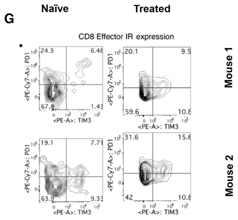
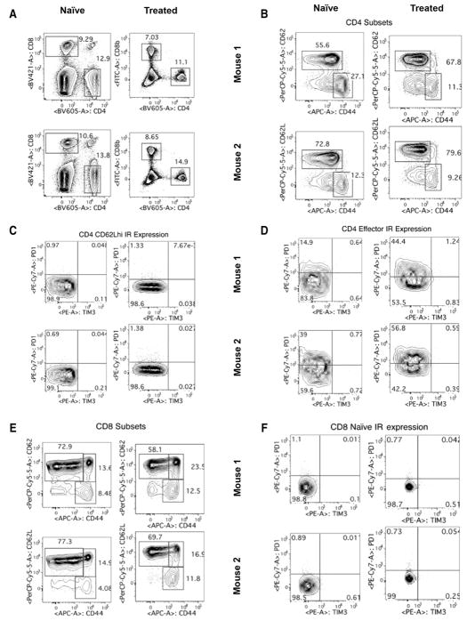
Phenotyping of T cells. Circulating lymphocytes from a tumor-naïve C57BL/6 mice (left column) were compared with those from C57BL/6 mice treated and cleared of B16 primary tumors (right column) (n = 2 mice/group, representative of four independent experiments). Multiparametric flow cytometry for live CD4+ or CD8+ T cells (A), the fraction of CD4+ or CD8+ cells that are CD62Lhi or effector (CD62Llo CD44hi) phenotype (B and E), the fraction of CD62Lhi CD4+ or CD8+ cells expressing the inhibitory receptors (IR) PD-1 and TIM-3, the fraction of CD62Llo CD44hi effector cells expressing the IRs PD-1 and TIM-3 (D and G). To analyze quantitative flow cytometry data, one-way ANOVA testing was conducted with a Tukey posttest; P values reported from these analyses were corrected to account for multiple comparisons.

