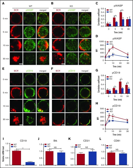
Figure 3.
The recruitment of pWASP and pCD19 to BCR clusters in B cells stimulated by sAg is decreased in Dock8 KO B cells. Murine splenic B cells were incubated with AF546–mB-Fab′–anti-Ig without or with streptavidin (sAg) at 4°C, washed, and warmed to 37°C for varying lengths of time. After fixation and permeabilization, the cells were stained for pWASP and pCD19 and analyzed using CFm (A-C,E-G) or flow cytometry (D,H). Flow cytometry analysis of the MFI of pWASP (D) and pCD19 (H) after stimulation with sAgs. The Pearson’s correlation coefficients between BCR and pWASP (C) or between BCR and pCD19 (G) staining in sAg-stimulated cells were determined using NIS-Elements AR 3.2 software. The relative mRNA levels of cd19, btk, cd21, and cd81 in splenic B cells examined by RT-PCR (I-L). Shown are representative images in which more than 50 cells were individually analyzed using NIS-Elements AR 3.2 software; shown are mean values (±SD) from 3 independent experiments. Scale bars, 2.5 μm. *P < .01; **P < .001.

