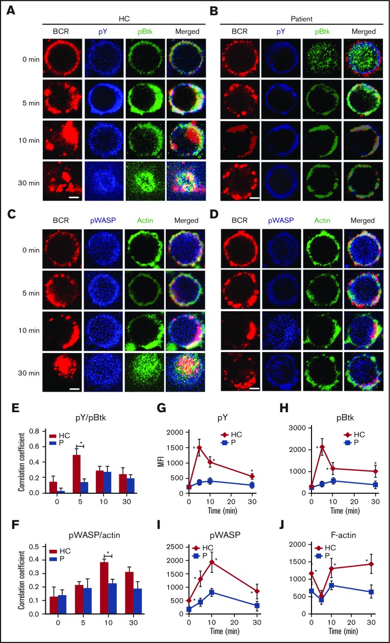
Figure 5.
The recruitment of BCR signalosomes and actin to BCR clusters is decreased in Dock8 patients. Human B cells from HCs and Dock8 patients were incubated with AF546–mB-Fab′–anti-Ig without or with streptavidin (sAg) at 4°C, washed, and warmed to 37°C for varying lengths of time. After fixation and permeabilization, the cells were stained for pY, pBtk, pWASP, and F-actin and were analyzed using CFm (A-D) or flow cytometry (G-J). The Pearson’s correlation coefficients between BCR and pY/pBtk (E) or between BCR and pWASP/actin (F) staining in sAg-stimulated cells were determined using NIS-Elements AR 3.2 software. Flow cytometry analysis of the MFI of pY (G), pBtk (H), pWASP (I), and F-actin (J) after stimulation with sAgs. Shown are representative images in which more than 50 cells were individually analyzed using NIS-Elements AR 3.2 software, and mean values (±SD) are from 3 independent experiments. All 3 patients were included, and each patient was an independent experiment. Scale bars, 2.5 μm. *P < .01. P, probability.

