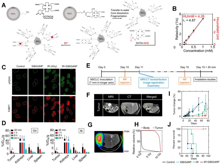
Figure 6.
Ultrasmall silica-based bismuth gadolinium nanoparticles (gadolinium-based AGuIX (Activation and Guidance of Irradiation by X-ray) nanoparticles with entrapped Bi (III)) for dual magnetic resonance/CT image-guided radiotherapy (IGRT). (A) These agents were synthesized by an original top-down process, which consists of Gd2O3 core formation, encapsulation by polysiloxane shell grafted with DOTAGA (1,4,7,10-tetraazacyclododecane-1-glutaric anhydride-4,7,10-triacetic acid) ligands, Gd2O3 core dissolution following chelation of Gd (III) by DOTAGA ligands, and polysiloxane fragmentation. Moreover, at the final stage of the synthesis, DOTA (1,4,7,10-tetraazacyclododecane-N,N',N'',N'''-tetraacetic acid)-NHS (N-hydroxysuccinimide) ligands were grafted to the surface to entrap free Bi3+ atoms into the final complex. (B) MRI (relaxivity) and CT (Hounsfield units) linear relation with concentration of nanoparticles (metal) in aqueous solution. (C) Qualitative representation of γ-H2AX and 53BP1 (p53-binding protein 1) foci formation, with and without 4 Gy irradiation, with and without nanoparticles, 15 min post-irradiation. (D) Biodistribution study performed by ICP-MS (inductively coupled plasma-mass spectrometry) in animals after intravenous injection of nanoparticles. (E) Experimental timeline based on a current clinical workflow for MRI-guided radiotherapy. (F) Fusion of the CT and MRI images. Yellow arrows indicate the inceased contrast in the tumor. (G) Dosimetry study performed for a single fraction of 10 Gy irradiation delivered from a clinical linear accelerator (6 MV). (H) Dose-volume histogram showing the radiation dose distribution in the tumor and in the rest of the body. (I) Mean tumor volume of each group. (J) Overall survival of each treatment cohort. Reproduced with permission from reference 207, copyright 2017 American Chemical Society.

