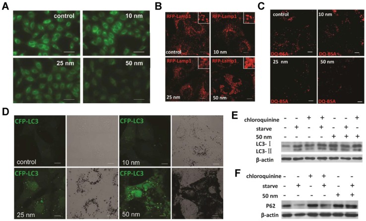
Figure 7.
AuNPs induce autophagosome accumulation through size-dependent nanoparticle uptake and lysosome impairment. (A) Effect of AuNPs on lysosome pH (representative fluorescence pictures of NRK cells (normal rat kidney) treated with AuNPs, then stained with LysoSensor Green DND-189 for evaluation of lysosomal acidity). Scale bar = 50 μm. (B) Vacuoles induced by AuNP treatment are enlarged lysosomes (inset: close-up of the enlarged lysosomes). Scale bar = 10 μm. (C) DQ-BSA (derivative-quenched bovine serum albumin, a self-quenched lysosome degradation indicator) analysis of lysosomal proteolytic activity. Accumulation of fluorescence signal, generated from lysosomal proteolysis of DQ-BSA, was much lower in AuNP-treated cells. Scale bar = 10 μm. (D) Formation of CFP (cyan fluorescent protein)-LC3 (microtubule-associated protein 1 light chain 3) dots (pseudocolored as green) in CFP-LC3 NRK cells treated with AuNPs. Left, confocal image; right, bright-field image. Scale bar = 10 μm. (E) LC3 turnover assay. The differences in LC3-I and LC3-II levels were compared by immunoblot analysis of cell lysates. (F) Degradation of the autophagy-specific substrate/polyubiquitin-binding protein p62/SQSTM1 (sequestosome 1) was detected by immunoblotting. Reproduced with permission from reference 280, copyright 2011 American Chemical Society.

