Figure 9.
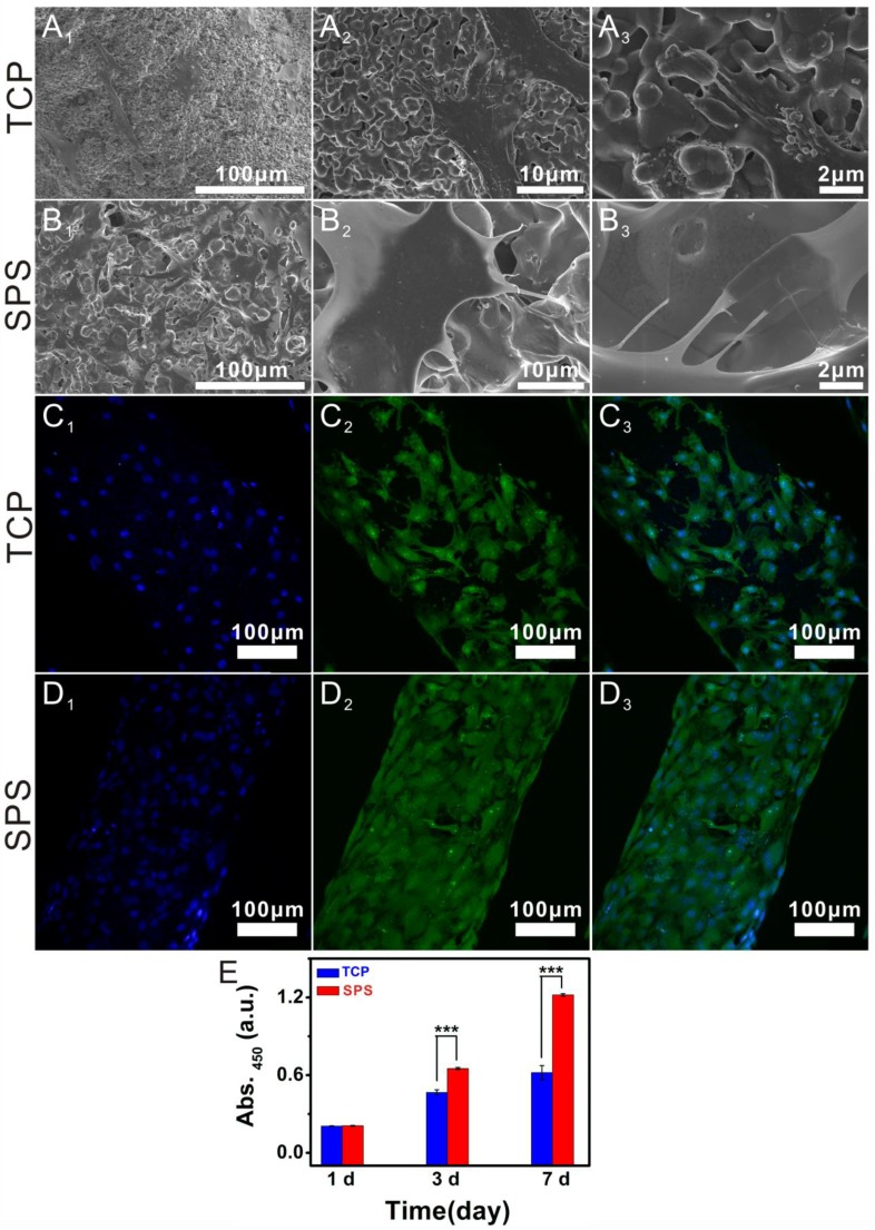
The attachment and morphology of chondrocytes cultured in SPS scaffolds. SEM images showed that chondrocytes attached to TCP scaffolds (A1-A3) and SPS scaffolds (B1-B3). CLSM images exhibited the morphology and cytoskeleton of chondrocytes cultured in TCP scaffolds (C1-C3) and SPS scaffolds (D1-D3). A1 and B1: 500×, A2 and B2: 3,000×, A3 and B3: 10,000×; C1 and D1: DAPI, C2 and D2: FITC, C3 and D3: merge images. (E) The proliferation of chondrocytes cultured in SPS and TCP scaffolds. Both TCP and SPS scaffolds supported the attachment of chondrocytes, and the chondrocytes in SPS scaffold exhibited better-defined microfilaments and cytoskeletons as compared to that of TCP scaffold. Compared with TCP scaffolds, SPS scaffolds distinctly elevated chondrocyte proliferation at 3 and 7 days. Repeat number: n=6. (*p<0.05, **p<0.01, ***p<0.001).

