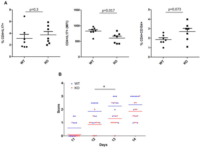
Figure 4.
Role of p70S6K1 in in vivo differentiation of MOG-specific Th17 cells and the onset of EAE. (A) Wild type and p70S6K1 knockout mice were immunized with MOG in complete Freund’s adjuvant (CFA) without administering pertussis toxin. Splenocytes from individual mice were harvested 10 days after immunization, and were cultured in the presence of MOG (20 μg/mL), IL-23 (20 ng/mL), and anti-IFN-γ antibody (10 μg/mL) for 3 days. These cultured splenocytes were stimulated with PMA/ionomycin for 4 h with last 2 h with monensin, and the percentage of CD4+ T cells produced IL-17 (left) was determined. The amount of IL-17 produced by CD4+ T cells (middle) was determined by measuring the MFI of CD4+IL17+ T cells. To measure the MOG-reactive CD4+ T cells (right), splenocytes were cultured overnight with monensin to determine the percentage of CD4+CD154+ T cells. p value was determined with Mann–Whitney test. (B) EAE was induced in WT and KO mice (10 mice/group) by immunizing with MOG35–55 peptide in CFA, followed by administration of pertussis toxin, and the animals were observed for disease progression (clinical score). Each symbol represents an individual animal and bars represent means. The differences in the neurological score along the disease progression were analyzed by two-way ANOVA followed by the Bonferroni post-hoc test. *p < 0.01.

