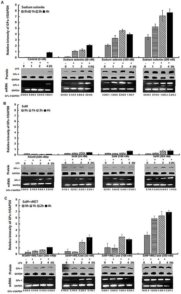
Figure 1.
GPx-1 expression in RAW264.7 macrophages. Cells were treated with 50nM, 100nM and 500nM Se-concentration in different forms: (A) Sodium selenite (SS), (B) Seleniferous wheat extract (SeW) and (C) Seleniferous wheat extract in presence of rMETase incubation (SeW+rMET) with respective to their controls, for 72 h and then inflamed with 1 µg/mL LPS for framed time interval upto 4 h. Densitometric values normalized to GAPDH are graphed with mean±S.D. for protein expression and indicated values below each panel of mRNA expression.

