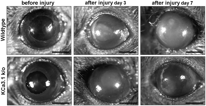
Fig 3. Loss of KCa3.1 reduced corneal haze in mice after alkali wounding.
Representative stereomicroscopic images showing corneal haze in Wild type (A, B, C), and KCa3.1-/- mice (D, E, F). Representative examples of Naïve (A, D), 3 days (B, E) and 7 days (C, F) alkali wounding are shown. A decrease in corneal haze was observed in KCa3.1-/- mice.

