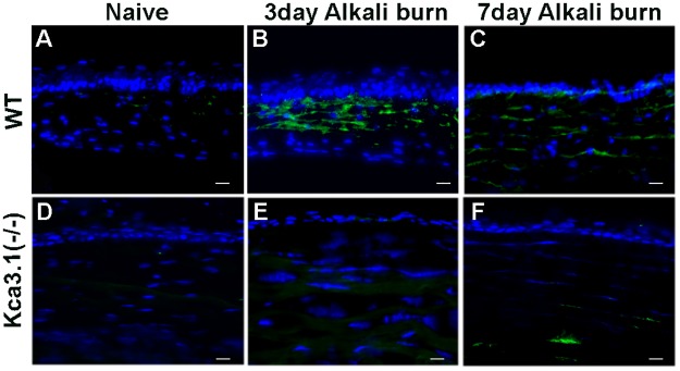
Fig 4. Effect of KCa3.1 loss on corneal fibrosis.
Corneal tissue immunostaining of mouse cornea showing levels of α-SMA expression in WT mice at 0, 3 and 7 days (A-C) and KCa3.1-/- mice at 0, 3 and 7 days (D-F) in alkali-induced corneal fibrosis. Blue: DAPI-stained nuclei and green: α-SMA staining. KCa3.1-/- mouse corneas showed a decrease in α-SMA expression in the stroma compared with that in control WT corneas.

