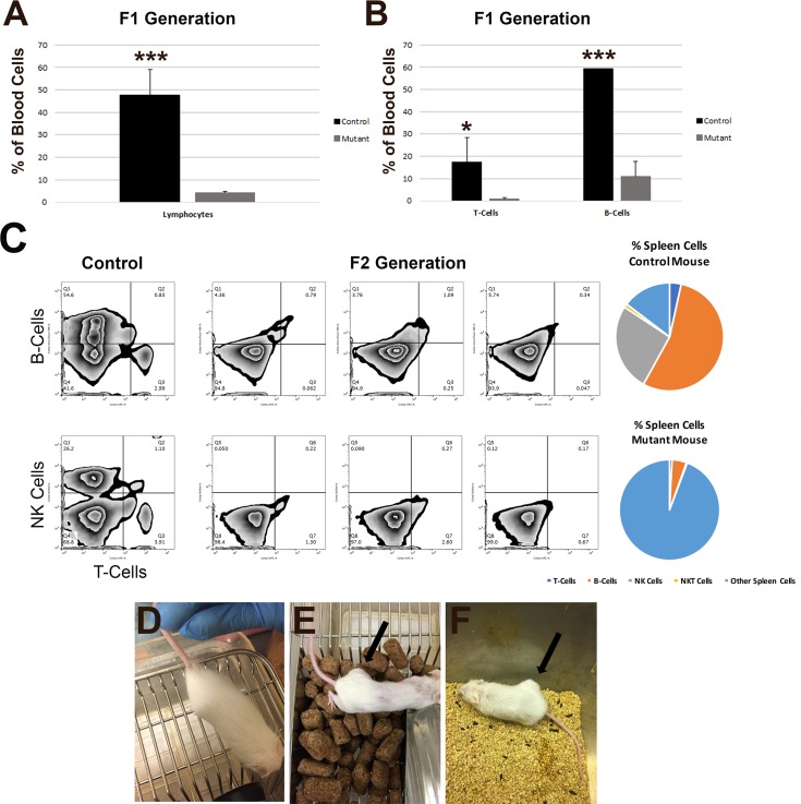
Fig 2. Immunocompromised state of the Rag2 and Il2rγ knockout Wsh/Wsh mice.
(A) Flow cytometry from blood for lymphocytes in Wsh/Wsh control mice and the first (F1) generation of Rag2 and Il2rγ mutant mice. (B) Flow cytometry from blood for the presence of T-cells and B-cells in Wsh/Wsh control mice and the F1 generation of Rag2 and Il2rγ mutant mice. *, p < 0.05; *** p < 0.001; N ≥ 2 control mice; N ≥ 4 mutant mice. (C) Flow cytometry of spleen cells in two control (far left) mice compared to three F2 generation Rag2 and Il2rγ mutant mice displaying no detectable populations of B-cells (top row, y-axis), T-cells (top and bottom rows, x-axis), or NK cells (bottom row, y-axis). Percentages of cell types from the flow cytometry analysis are shown for both the control mouse (top right) and mutant moue (bottom right). (D-F) MCF7 human mammary carcinoma cells were injected subcutaneously into the flank of four Wsh/Wsh mice (D), an immune-compromised NOD SCID gamma mouse (E), and five Wsh/Wsh Rag2 and Il2rγ mutant mice (F). Human MCF7 cells survived and proliferated only in the immunocompromised NOD SCID gamma mouse and Wsh/Wsh Rag2 and Il2rγ mutant mice after 6 weeks post-injection. No human cells displayed growth and proliferation in Wsh/Wsh mice by 7 weeks post-injection. Arrows, tumor-like growth sites.

