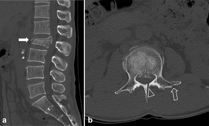
Figure 3.

Low-dose CT images of a 48-year-old male with a history of falling down. The body mass index and effective dose were 20.9 kg m–2 (lower range of normal-weight, body mass indexGroup 1) and 1.2 mSv, respectively. Sagittal (a) and axial (b) CT images show acute fractures of the body (burst fracture, white arrow, a), left transverse process (open arrow, b) and lamina (open arrowhead, b) of the L2. Both reviewers assigned a score of 1 for diagnostic acceptability (fully acceptable) and diagnosed the fractures correctly.
