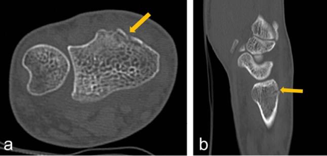
Figure 3.

A 45-year-old male with right wrist pain after a fall. He had tenderness, swelling of the joint and limited movement. Plain radiography did not show definite fracture lines. (a) Axial image of standard-dose CT scan showing faint cortical disruption (arrow). (b) Sagittal reconstructed image showing cortical disruption (arrow). The patient was diagnosed with an avulsion fracture of the distal radius clinically and radiologically.
