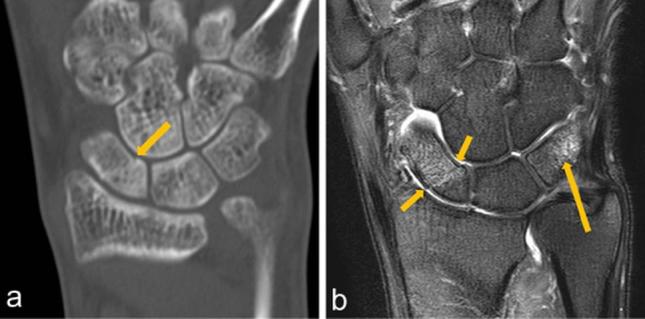
Figure 4.

A 37-year-old male with left hand pain after a fall. He had tenderness and swelling of the hand. (a) Coronal image of low-dose CT scan showing faint cortical disruption (arrow) in the proximal scaphoid. This fracture was misdiagnosed as normal by one of interpreters. (b) Immediate follow-up MRI. Coronal T2 weighted fat-suppressed image (TR/TE, 4000 ms/90 ms) at the same portion revealed fracture lines and combined bone marrow edema (small arrows). Bone marrow edema was also evident in the triquetrum (long arrow). No apparent fracture line was seen on the CT even in the second evaluation.
