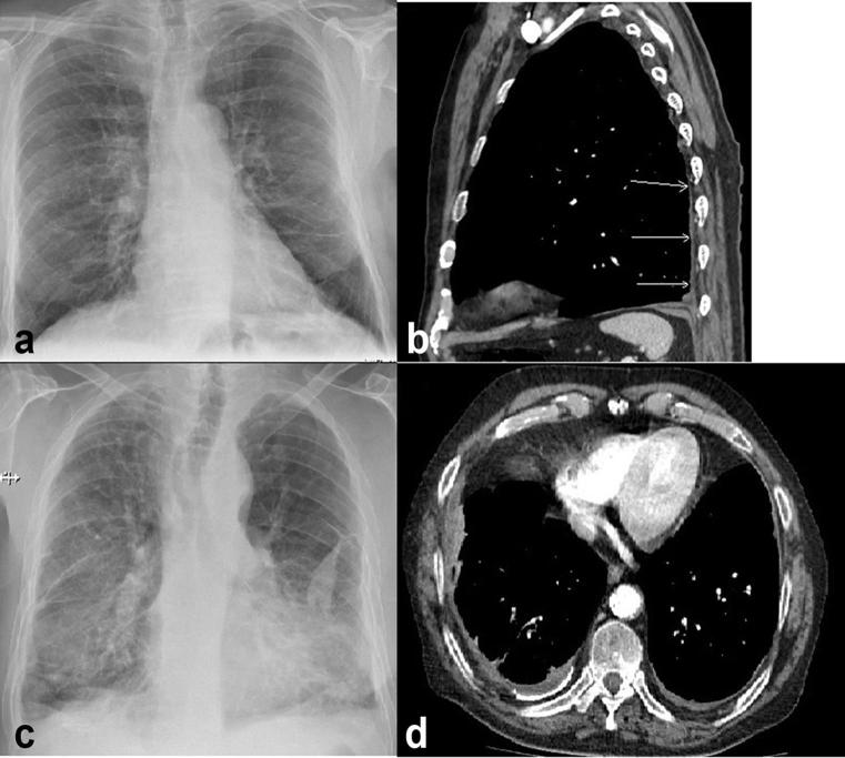
Figure 2.
(a) Postero-anterior chest radiograph of a patient with right-sided diffuse pleural thickening (DPT) with no costophrenic angle obliteration. (b) Sagittal CT view of the same patient showing DPT involving the posterior pleura. (c) Patient with bilateral DPT and costophrenic angle obliteration. (d) Axial CT image confirming bilateral pleural thickening.

