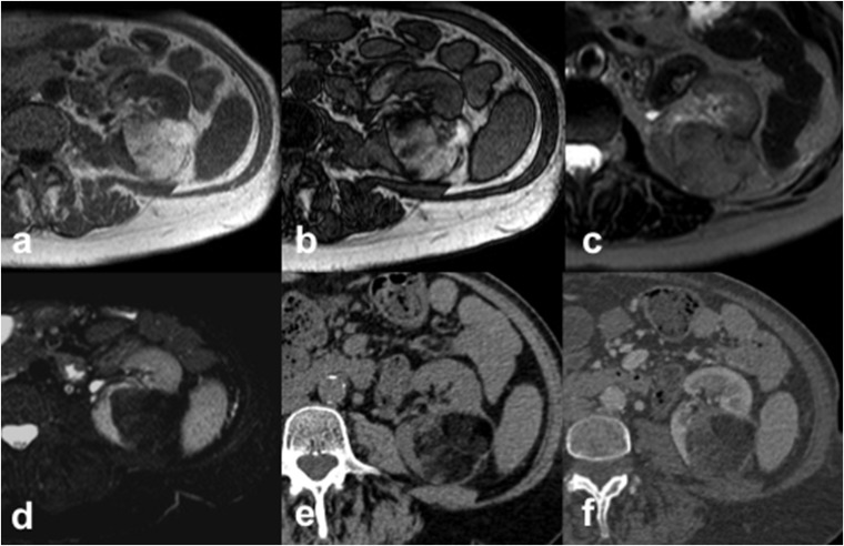
Figure 1.
Angiomyolipoma (AML) in a 63-year-old female. Axial T1 weighted in-phase (a) and out-of-phase (b) gradient recalled echo images show an hyperintense left-renal AML with the presence of “india ink artefact”, seen as an interface between the AML and the kidney. The AML shows intermediate signal intensity on axial T2 weighted fast spin echo image (c) with a loss of signal on axial T2 weighted fat-saturated image (d). Axial CT images acquired before (e) and after (f) intravenous contrast administration demonstrate the typical features of AML appearing as a fat-containing renal lesion with no calcifications.

