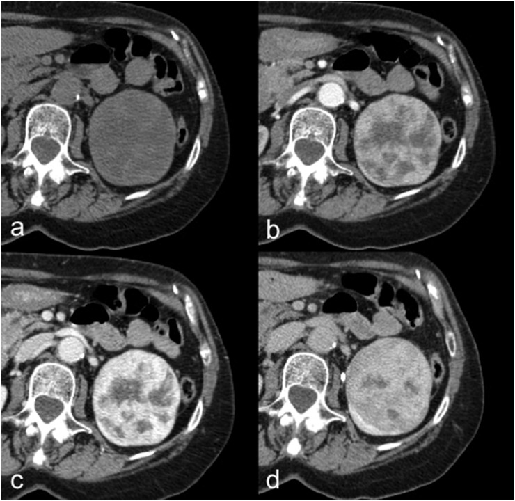
Figure 3.
Oncocytoma in a 72-year-old female. Axial unenhanced (a), corticomedullary phase (b), nephrographic phase (c) and excretory phase (d) CT images show a well-circumscribed left-renal lesion with inhomogeneous enhancement and a central stellate scar, without calcifications, necrosis or haemorrhage.

