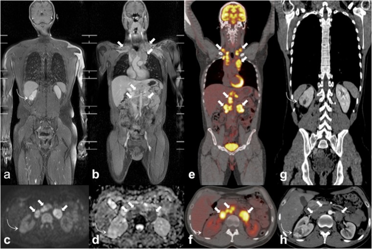
Figure 5.
Incidentally discovered clear cell renal cell carcinoma (ccRCC) during the staging work-up of a 35-year-old male patient with Hodgkin lymphoma. Coronal whole-body MR three-dimensional fat-suppressed T1 weighted gradient recalled echo images after paramagnetic contrast administration (a, b) show an incidental right renal mass (curved arrows) and multiple enlarged lymph nodes (arrows) in bilateral supraclavicular, mediastinal and retroperitoneal regions. On axial high b-value diffusion-weighted imaging image (c) and axial apparent diffusion coefficient map (d), the ccRCC (curved arrows) does not show the restricted pattern of diffusion demonstrated by lymphomatous nodes (arrows). The ccRCC (curved arrow) also shows poor fluorine-18 fludeoxyglucose (18F-FDG) uptake on axial 18F-FDG positron emission tomography (PET)/CT (f) in respect of the locations of lymphoma (arrows) that are markedly 18F-FDG avid on coronal (e) and axial (f) 18F-FDG-PET/CT images. Coronal (g) and axial (h) portal-phase CT images demonstrate the high and inhomogeneous enhancement of the ccRCC (curved arrows) and the homogeneous and low enhancement of lymphoma (arrows).

