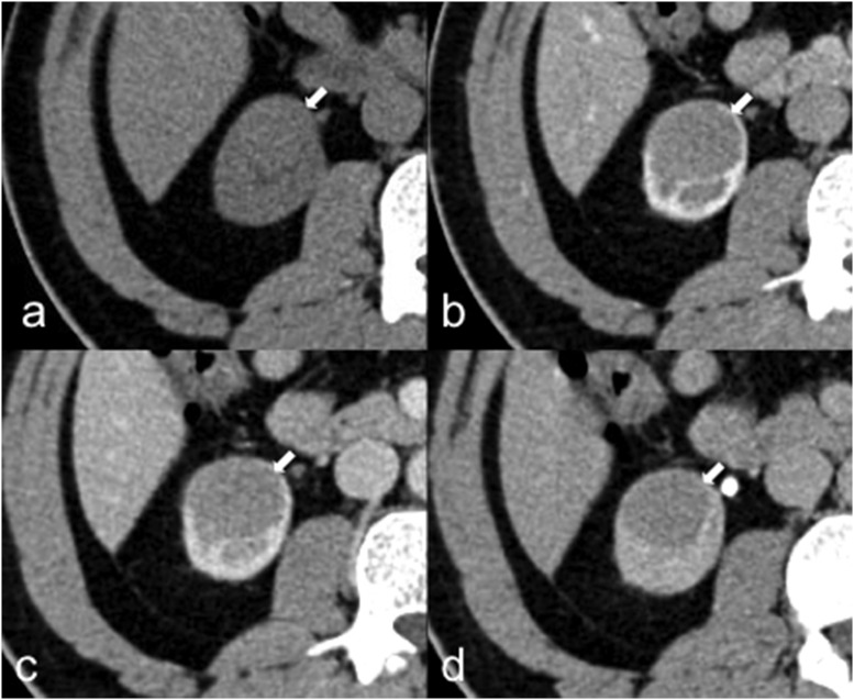
Figure 7.
A 40-year-old male with papillary renal cell carcinoma (pRCC). Axial unenhanced (a), corticomedullary phase (b), nephrographic phase (c) and excretory phase (d) CT images demonstrate a right-renal homogeneous mass (arrows) with poor and slow enhancement, typically observed in pRCC that is hypovascular in comparison with clear cell renal cell carcinoma.

