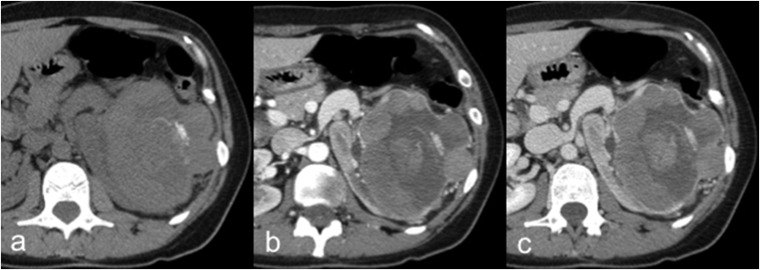
Figure 8.
A 45-year-old female with chromophobe renal cell carcinoma (cRCC). Axial unenhanced (a), corticomedullary phase (b) and nephrographic phase (c) CT images show a large hypovascular and inhomogeneous left renal mass with intratumoural calcifications and necrotic areas. The cRCC usually demonstrates lower post-contrastographic enhancement than clear cell renal cell carcinoma.

