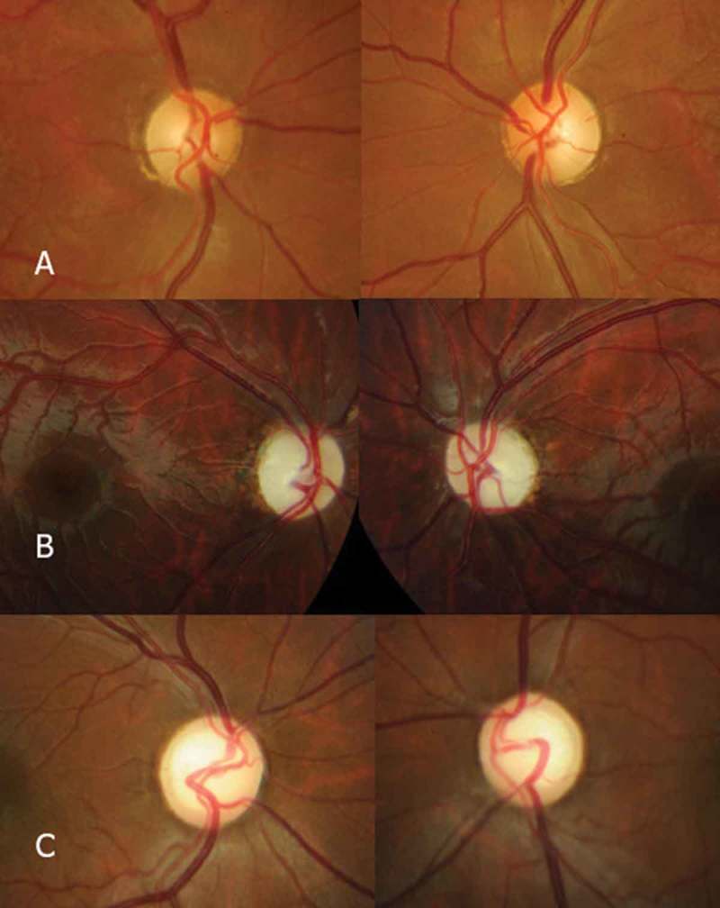
Figure 1.

Photocomposition showing the appearance of the optic nerves of Patients 3 (A), 4 (B), and 12 (C) among the WFS+ patients. Patient 3 exhibited optic nerve pallor localised in the temporal sector, which occurred in 80.5% of the patients. Patient 2 showed global pallor of the optic nerve, which occurred in 19.4% of the patients. Finally, Patient 12 exhibited pallor and excavation, which occurred in 22.2% of the patients.
